

In October 2021, during the opening of Panzhihua Daguanming Eye Clinic, they introduced Big Vision Medical’s fully automatic AI OCT BV1000, becoming the first optometry clinic in China to introduce AI OCT and conduct comprehensive OCT examination services. After more than nine months of practical application, the founder and chairman of Panzhihua Daguanming Eye Clinic, Xue Kejun, has gained a more personal understanding of the high-tech attributes of this AI OCT machine. Recently, Xue Kejun shared his experience of using the equipment and expressed a positive outlook for the application of advanced AI technology in grassroots ophthalmology.

Xue Kejun, the chairman, first told us that “introducing Big Vision Medical’s AI OCT is an absolutely correct choice for optometry clinics in transformation and upgrading.” Many optometry centers or clinics, either prior to or after transformation, continue to mainly focus on optometry and glasses fitting. Their professional level is relatively weak, and although they understand the importance of OCT in ophthalmic examination, it is difficult to conduct OCT examinations without expertise. Some optometry centers and clinics with strong capabilities also have low utilization rates of traditional OCT machines. With advanced ophthalmic AI technology that achieves accuracy comparable to that of chief physicians, these difficulties can be easily overcome, making it possible to actively carry out many services that were previously impossible.

For optometry centers and clinics, the main target population is myopic patients, and a considerable proportion of them are highly myopic. According to information released by the National Health Commission, in 2020, nearly 10% of myopic students nationwide were highly myopic, and the proportion increased with grade level; 1.5% of 6-year-old children in kindergarten were highly myopic, while the percentage in high school reached 17.6%. The risk of fundus lesions in highly myopic patients is much higher than that of ordinary people, and highly myopic patients should undergo regular fundus examinations. These are common knowledge for optometry practitioners, but in practice, it is difficult to carry out regular check-ups and follow-up visits for highly myopic patients.
Generally speaking, fundus lesions are a relatively long-term process. Large hospitals have a large number of patients, and examinations require queuing, so most highly myopic patients are not aware of the need for regular check-ups in hospitals. When they go to optometry centers or clinics for refraction and fitting, these centers or clinics often lack professional fundus examination capabilities. Taking retinal detachment, a frequent complication in highly myopic patients, as an example, the retinal tears located beside the blood vessels are often in the stationary stage and do not progress further. In this case, only observation and follow-up of the progress of the condition are required. For retinal tears that continue to expand or eventually lead to retinal detachment, early intervention or surgical treatment is necessary. In any case, strict follow-up and observation are crucial. In practice, the proportion of highly myopic patients who undergo regular fundus examinations in China is very low. The “14th Five-Year” National Eye Health Plan proposed earlier this year aims to gradually reduce the number of vision loss caused by high myopia. An effective way to achieve this goal is to enhance the fundus examination service capabilities of optometry centers and clinics, and advanced intelligent ophthalmic AI OCT can play an important role in this work.
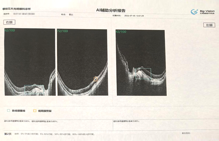
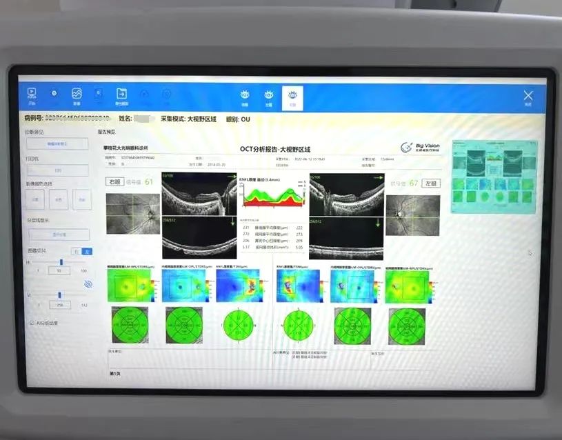
AI OCT clearly shows the presence of choroidal atrophy and retinal tears.
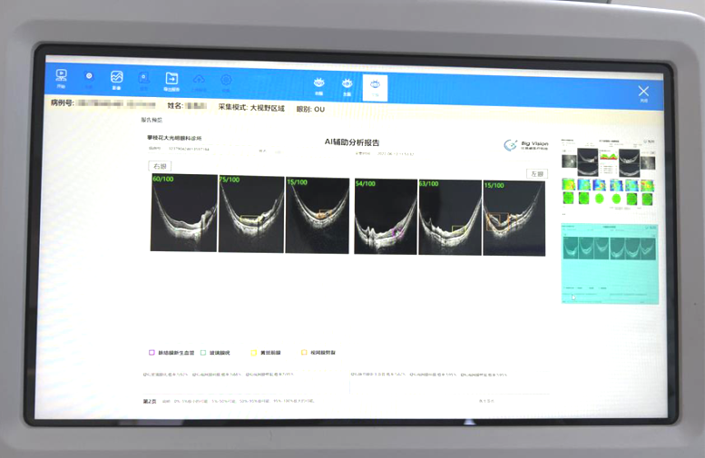
AI OCT showed a more severe degree of fundus lesions.
Myopia prevention and control
Given the high myopia rate among Chinese adolescents and the increasing demand for vision improvement,
Orthokeratology fitting has developed rapidly in recent years and has become a major business growth point for optometry centers and clinics. According to the “Expert Consensus on Orthokeratology Fitting Process” (2021) released by the Eye Optometry Group of the Ophthalmology Branch of the Chinese Medical Association, comprehensive pre-fitting examinations should be performed before orthokeratology fitting, including naked eye visual acuity, optometry, corneal curvature, corneal topography, intraocular pressure, fundus, and axial length. Among these items, fundus examination is relatively unfamiliar to optometry centers and clinics, and requires a high degree of expertise, which has become an important factor limiting the implementation of orthokeratology fitting. With the assistance of AI-enabled OCT examination, optometrists can have a comprehensive understanding of the patient’s retinal condition before fitting, and can also perform regular fundus examinations during the wearing period, thereby ensuring the fitting and lifelong management of orthokeratology lenses.
Chairman Xue has personally experienced missing the optimal treatment window due to late detection of fundus disease, and has a firsthand understanding of the visual damage caused by fundus disease. Therefore, he has always instilled in the company’s operation a philosophy that no eye disease patient should slip away from us, and patients must receive standardized treatment in a timely manner.
In the past, due to the limited development of industry technology, optometry centers and clinics had insufficient ability to detect diseases, making it very difficult to achieve this goal. With advanced ophthalmic AI technology, optometry centers and clinics are now able to judge referrals, establish cooperative relationships with professional ophthalmic hospitals, and achieve graded diagnosis and treatment through division of labor and collaboration. This is beneficial for patients, optometry centers or clinics, and professional ophthalmic hospitals, and strengthens the connection between optometry centers and clinics and professional ophthalmic hospitals.

What also pleases Chairman Xue is that with the help of OCT images, many users see their retinal structure for the first time, gaining a more intuitive understanding of eye structure and increasing awareness of eye health. It makes everyone responsible for their own eye health, which is crucial to the promotion of eye health. Meanwhile, Chairman Xue is actively recommending the introduction of AI-OCT to other optometry colleagues. As a veteran who has been working in the field of optometry for decades, he is happy to see the technological progress in the industry and embrace emerging AI technology, which has once again given him great motivation for the eye optometry industry.


No comments yet.