

Recently, Big Vision, in collaboration with ophthalmology experts Director Peng Qing and Director Gao Peng from the Shanghai Tenth People’s Hospital, published a paper in the renowned journal Frontiers in Cell and Development Biology (impact factor: 6.081). This study verified the feasibility and effectiveness of Big Vision’s ophthalmic AI in community eye disease screening.
Age-related macular degeneration (AMD), diabetic retinopathy (DR), high myopia, as well as macular diseases such as macular epiretinal membrane and macular hole are important factors causing vision loss. Early diagnosis and timely treatment of these eye diseases are critical to achieving the best visual outcomes. Therefore, screening in primary hospitals and communities plays an important role.

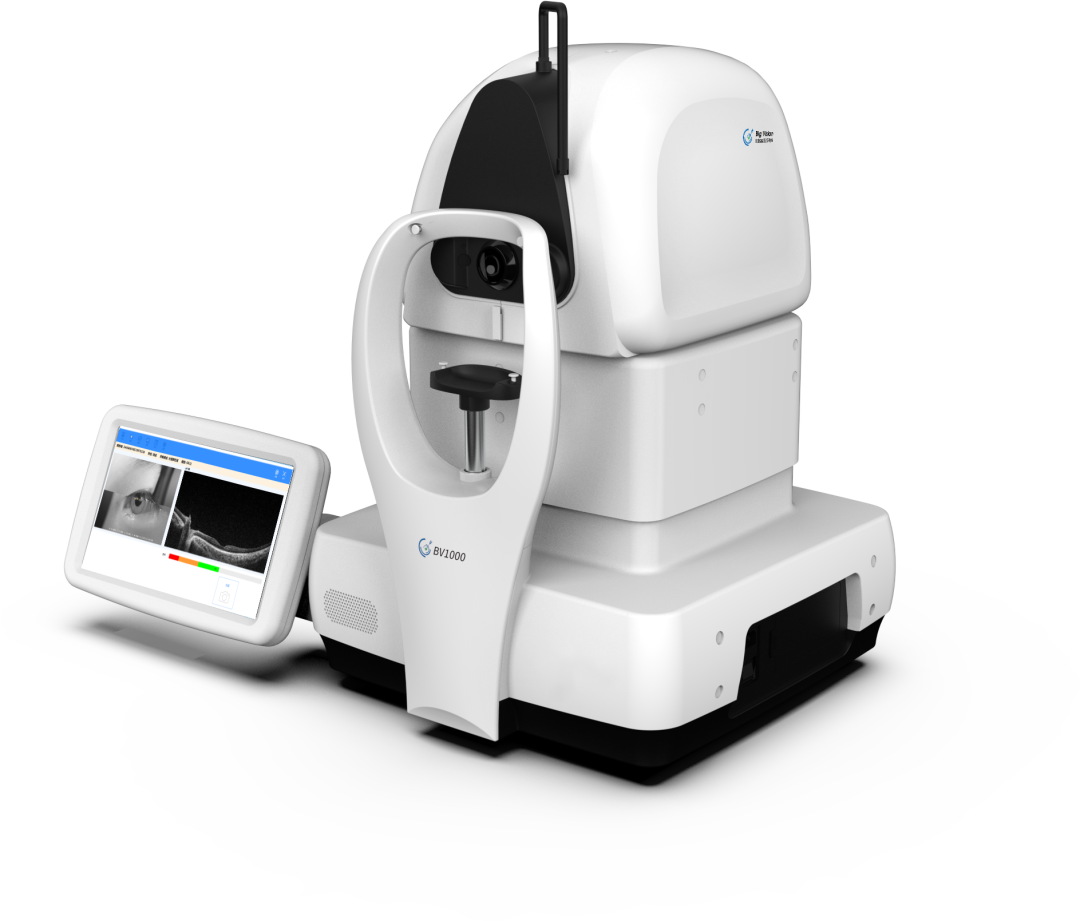
In recent years, there has been a contradiction between the shortage of senior ophthalmologists in primary community hospitals and the strong demand for screening, as well as some defects found in the screening process. Based on optical coherence tomography (OCT) imaging using the BV1000 scanner, Big Vision Medical Technology applies AI algorithms to analyze the retinal layers, especially in the macular area, and detect various retinal diseases, which can significantly improve the screening capabilities of primary community hospitals for eye diseases.


Abstract
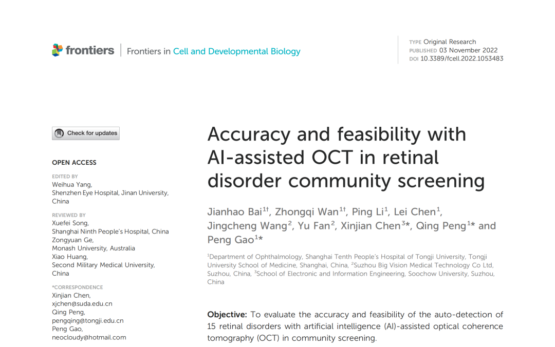
Objective:
To evaluate the accuracy and feasibility of an artificial intelligence (AI)-assisted optical coherence tomography (OCT) system in automatically detecting 15 kinds of retinal diseases during community screening.
Methods and Discussions:
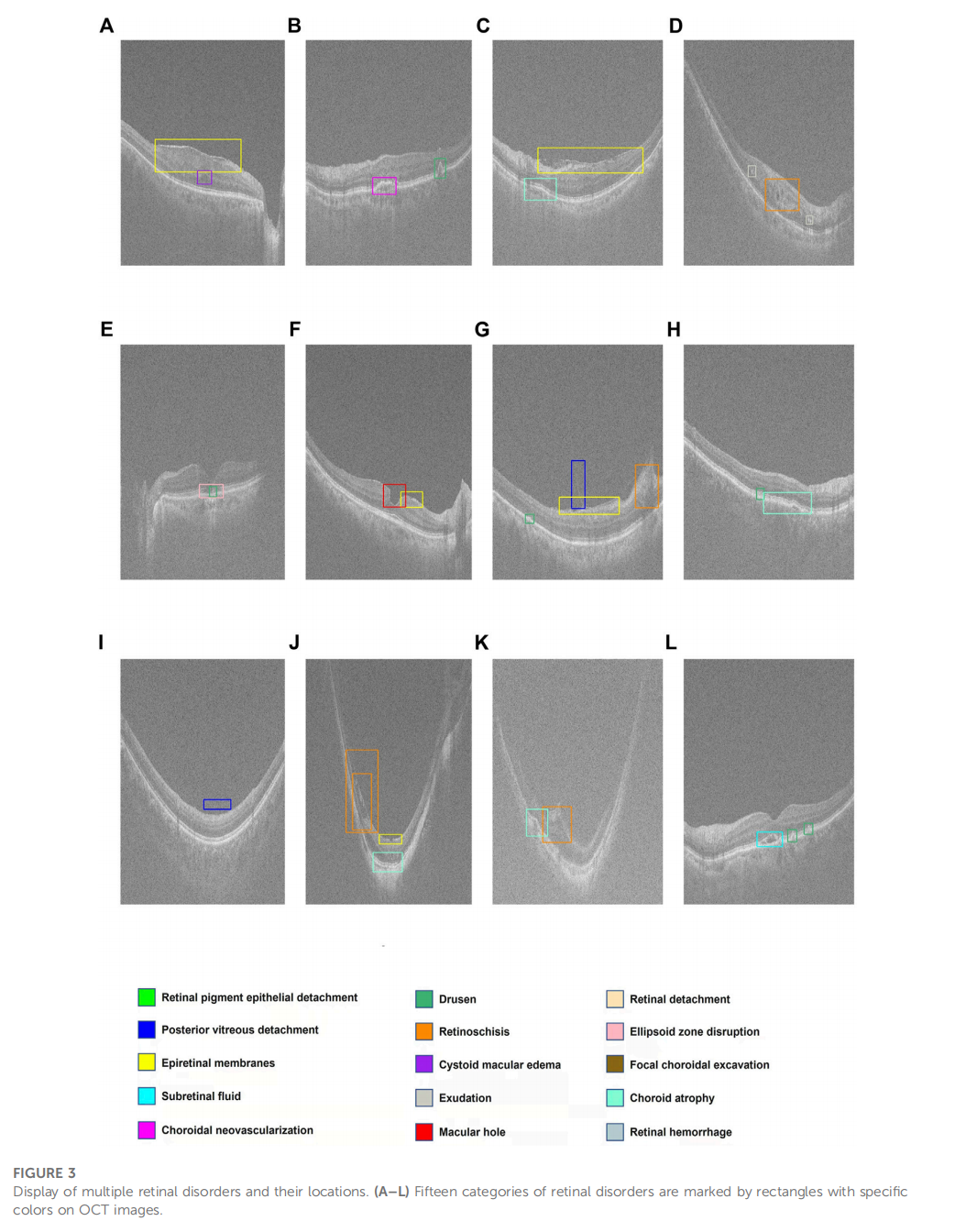
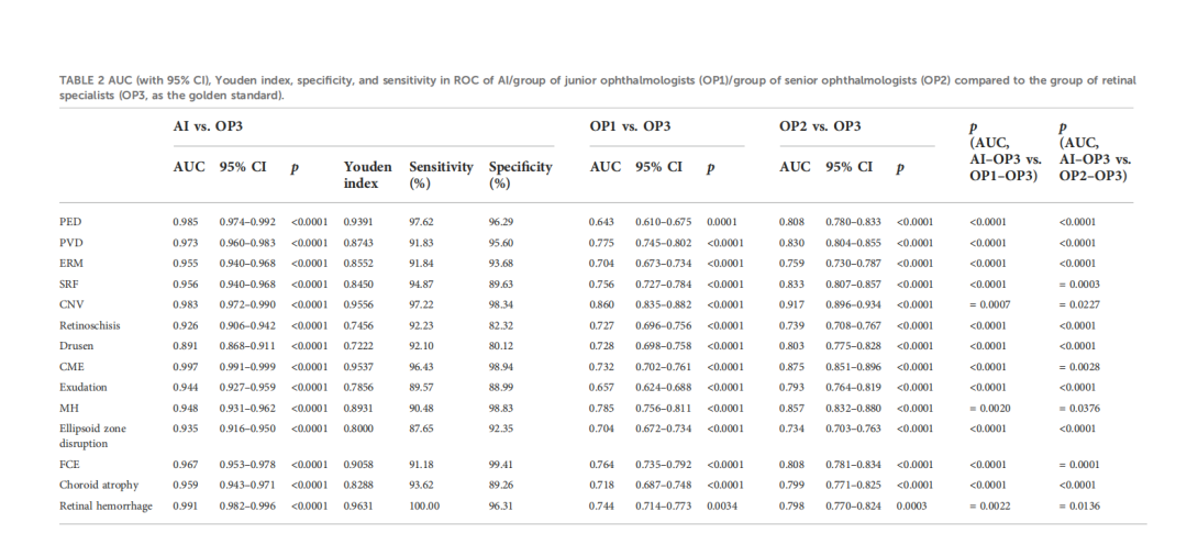
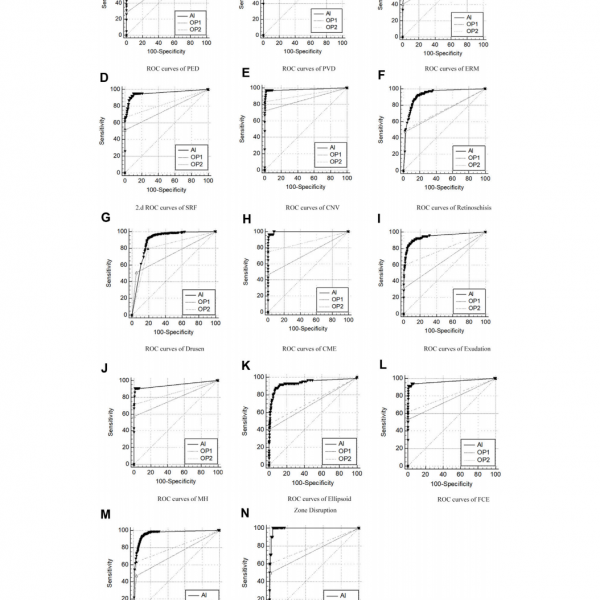
From September to December 2021, 477 subjects from four communities, with a total of 954 eyes, were included in this study. The macular disc area of the posterior pole of the subjects’ retina was captured using the wide-field mode (12mm x 9mm) of the BV1000 scanner, and ophthalmic examinations were performed in conjunction with the subjects’ basic information. An integrated software and deep-learning algorithm were used to analyze the OCT images, enabling detection of 15 kinds of retinal diseases, including pigment epithelial detachment (PED), posterior vitreous detachment (PVD), epiretinal membrane (ERM), subretinal fluid (SRF), choroidal neovascularization (CNV), vitreomacular traction, retinal schisis, cystoid macular edema (CME), exudation, macular hole (MH), retinal detachment (RD), ellipsoid zone loss, focal choroidal excavation (FCE), choroidal atrophy, and retinal hemorrhage.
Meanwhile, three groups of ophthalmologists (retina specialists, senior ophthalmologists, and junior ophthalmologists) independently completed the diagnosis of the same group of subjects and compared their results with those of AI. The differences between the groups of doctors and AI were analyzed by calculating the area under the ROC curve (AUC), sensitivity, and specificity, and kappa statistics were performed. Results: Finally, 878 eyes were included, of which 76 were excluded due to poor image quality. In the detection of 15 retinal diseases, the AI exhibited a larger AUC (0.891-0.997), high sensitivity (87.65-100%), and high specificity (80.12-99.41%) compared to the results of the retina specialists. Compared with retina specialists, senior ophthalmologists, and junior ophthalmologists, AI was closest to the results of the retina specialists (p < 0.05).
Conclusion
AI-assisted OCT detection has high accuracy, sensitivity, and specificity in the automatic detection of 15 retinal diseases, demonstrating its feasibility and effectiveness in community ophthalmic screening.


No comments yet.